��������������������������
EXTERNAL HOOF
ANATOMY 101
PART 2
��������������������������������������������������������������������������������������������������
To repeat, nature made some clever engineering designs when the
hoof was created. Still not 100% understood, it takes a beating with
every step, undergoes stress levels that are off the chart and still
manages to move the horse forward. Part two of this series examines
the internal structures of the hoof and how involved and dependent
each structure is to allow the equine movement we take for granted.
Putting the anatomy into a 3D mindset is a good start to understanding
the inner hoof and the system of pulleys and levers that make it operate.
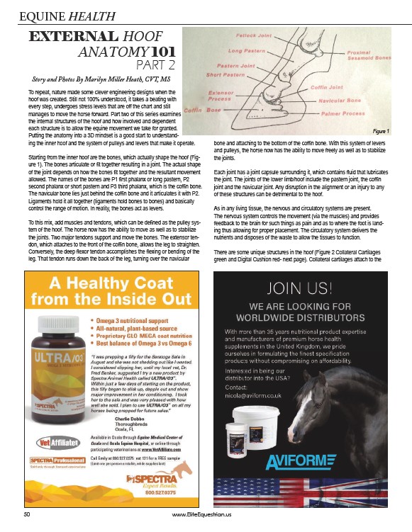
Starting from the inner hoof are the bones, which actually shape the hoof (Figure
1). The bones articulate or fit together resulting in a joint. The actual shape
of the joint depends on how the bones fit together and the resultant movement
allowed. The names of the bones are P1 first phalanx or long pastern, P2
second phalanx or short pastern and P3 third phalanx, which is the coffin bone.
The navicular bone lies just behind the coffin bone and it articulates it with P2.
Ligaments hold it all together (ligaments hold bones to bones) and basically
control the range of motion. In reality, the bones act as levers.
To this mix, add muscles and tendons, which can be defined as the pulley system
of the hoof. The horse now has the ability to move as well as to stabilize
the joints. Two major tendons support and move the bones. The extensor tendon,
which attaches to the front of the coffin bone, allows the leg to straighten.
Conversely, the deep flexor tendon accomplishes the flexing or bending of the
leg. That tendon runs down the back of the leg, turning over the navicular
bone and attaching to the bottom of the coffin bone. With this system of levers
and pulleys, the horse now has the ability to move freely as well as to stabilize
the joints.
Each joint has a joint capsule surrounding it, which contains fluid that lubricates
the joint. The joints of the lower limb/hoof include the pastern joint, the coffin
joint and the navicular joint. Any disruption in the alignment or an injury to any
of these structures can be detrimental to the hoof.
As in any living tissue, the nervous and circulatory systems are present.
The nervous system controls the movement (via the muscles) and provides
feedback to the brain for such things as pain and as to where the foot is landing
thus allowing for proper placement. The circulatory system delivers the
nutrients and disposes of the waste to allow the tissues to function.
There are some unique structures in the hoof (Figure 2 Collateral Cartilages
green and Digital Cushion red- next page). Collateral cartilages attach to the
50 www.EliteEquestrian.us
Figure 1