Sepideh Mokhtari, MD
Dr. Sepideh Mokhtari
is a neuro-oncologist
interested in the
neurologic complications
of cancer and
its treatment, as well
as neuro-oncology.
She did her neurology
training at Houston
Methodist and two
years of neuro-oncology fellowship at
Memorial Sloan Kettering Cancer Center
(MSKCC). She has special interests in
immune-mediated neurologic diseases as a
result of cancer or immune therapy. She has
been applying her experience from MSKC to
provide the best care to patients at Moffitt
Cancer Center. Since starting at Moffitt in
August 2016, she has been the director of
Neurological Services and associate director
of the Neurofibromatosis Clinic.
Dr. Mokhtari has her own personal story
about Moffitt’s outstanding physicians and
team members, all of whom truly value
patient-centered care. Soon after she
joined Moffitt, she entrusted the care of a
family member to Moffitt’s highly trained
neurosurgeon, Nam Tran, MD, PhD, for a
very high-risk operation. Dr. Mokhtari’s
brother-in-law is a 49 year old man with
long history of multiple sclerosis (MS)
dating back to 2000. Ten years ago, he
began losing strength on his right side.
This led to an MRI of the cervical spine,
which identified an enhancing cervical
cord lesion related to his MS. His weakness
continued to progress despite multiple
treatments for his MS, leading to lost
strength in his left leg and, eventually,
his left hand. This progressive, debilitating
weakness led to the diagnosis of secondary
progressive MS. Due to the complexity of
the situation, his case was presented at
multiple top cancer centers across the
nation. Some attributed his cervical cord
lesion to a benign neoplasm unrelated to
his MS. Multiple surgeons refused to
operate on him due to the high risk of
complete paralysis.
His clinical course continued to get worse,
resulting in severe spasticity and pain.
The most recent MRI of his cervical spine
showed progression of the lesion over the
last two years. Dr. Mokhtari discussed the
case of her brother-in-law with Dr. Tran.
After reviewing all the scans, Dr. Tran was
confident the lesion in his cervical spine
continues on page 5
EEG in Neuro-Oncology
By Edwin Peguero, MD
Since its first use
in humans in 1924,
electroencephalography
(EEG) has been
instrumental in the
diagnosis of epilepsy
and encephalopathy.
Special electrodes
attached to the scalp
in a standardized way
are used to measure the electrical potential
between two anatomical points in the
scalp. These spatial arrays, or montages,
allow us to identify and localize electrical
abnormalities, which in turn will help
develop independent management of
different clinical entities such as seizures
and encephalopathies.
The latter part is important in order to
achieve the successful diagnosis and
treatment of the patient.
presentations, can be challenging to detect
through standard electroencephalography.
However, with new modalities such as
72-hour video EEG ambulatory as well as
inpatient long-term monitoring, a more
accurate diagnosis can be made. This is
epitomized in the nonresponsive patient
who is critically ill, as they have a high
incidence of subclinical seizures. Our
group is actively participating in the
introduction of these tools into the
realm of novel treatments such as in
CAR T-cell therapy along with immuno-
therapy and standard chemotherapy.
Newer software has facilitated the
analysis of electroencephalographic
activity (i.e. spike detection systems,
ictal seizure detection, suppression/
asymmetry of the hemispheres,
artifact suppression/filtering).
This field is continuing to evolve
into a user-friendly tool with very
helpful clinical implications.
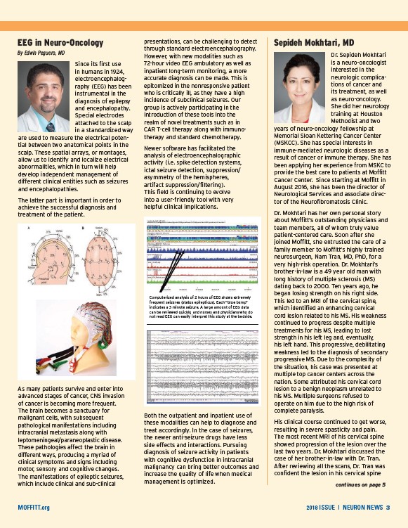
Computerized analysis of 2 hours of EEG shows extremely
frequent seizures (status epilepticus). Each “blue bump”
indicates a 3 minute seizure. A large amount of EEG data
can be reviewed quickly, and nurses and physicians who do
not read EEG can easily interpret this study at the bedside.
As many patients survive and enter into
advanced stages of cancer, CNS invasion
of cancer is becoming more frequent.
The brain becomes a sanctuary for
malignant cells, with subsequent
pathological manifestations including
intracranial metastasis along with
leptomeningeal/paraneoplastic disease.
These pathologies affect the brain in
different ways, producing a myriad of
clinical symptoms and signs including
motor, sensory and cognitive changes.
The manifestations of epileptic seizures,
which include clinical and sub-clinical
Both the outpatient and inpatient use of
these modalities can help to diagnose and
treat accordingly. In the case of seizures,
the newer anti-seizure drugs have less
side effects and interactions. Pursuing
diagnosis of seizure activity in patients
with cognitive dysfunction in intracranial
malignancy can bring better outcomes and
increase the quality of life when medical
management is optimized.
MOFFITT.org 2018 ISSUE | NEURON NEWS 3
/MOFFITT.org